An interview with Kareem Elsayad
A series of Euro-BioImaging Ambassador program – Introducing the Austrian BioImaging/CMI Node.
If you want to understand the physical properties of biological samples, Euro-BioImaging’s Austria BioImaging/CMI Node in Vienna might be a useful contact point for your research. Kareem Elsayad, a group leader at the Medical University of Vienna, took our Scientific Ambassador, Magdalena Schindler, on a tour of the range of Brillouin Light Scattering (BLS) based spectroscopy and microscopy techniques that he is developing with his group.
In this interview, Kareem explains how they are applying these techniques to better understand different biological systems and processes. They are especially open for collaborative multidisciplinary projects bridging the life sciences and physics, with access to their instruments and services also possible through the Austrian Node of Euro-BioImaging.
Understanding mechanics on the microscopic-scale at the Austrian BioImaging/CMI with Brillouin Light Scattering Microscopy

The research of the Elsayad Lab at the Center for Anatomy and Cell Biology of the Medical University of Vienna focuses for the most part on developing and applying novel optical microscopy and spectroscopy techniques to explore mechanical, structural and dynamic properties of biological materials at the microscopic scale. Their overarching goal is to understand how these properties can give rise to and play a role in fundamental biological processes as well as how they might be affected in diseases. For the latter case they also have a keen interest in establishing the developed techniques as routine R&D tools in biomedical research and translating them to the clinic where they could form the basis of novel prognostic and diagnostic methods.
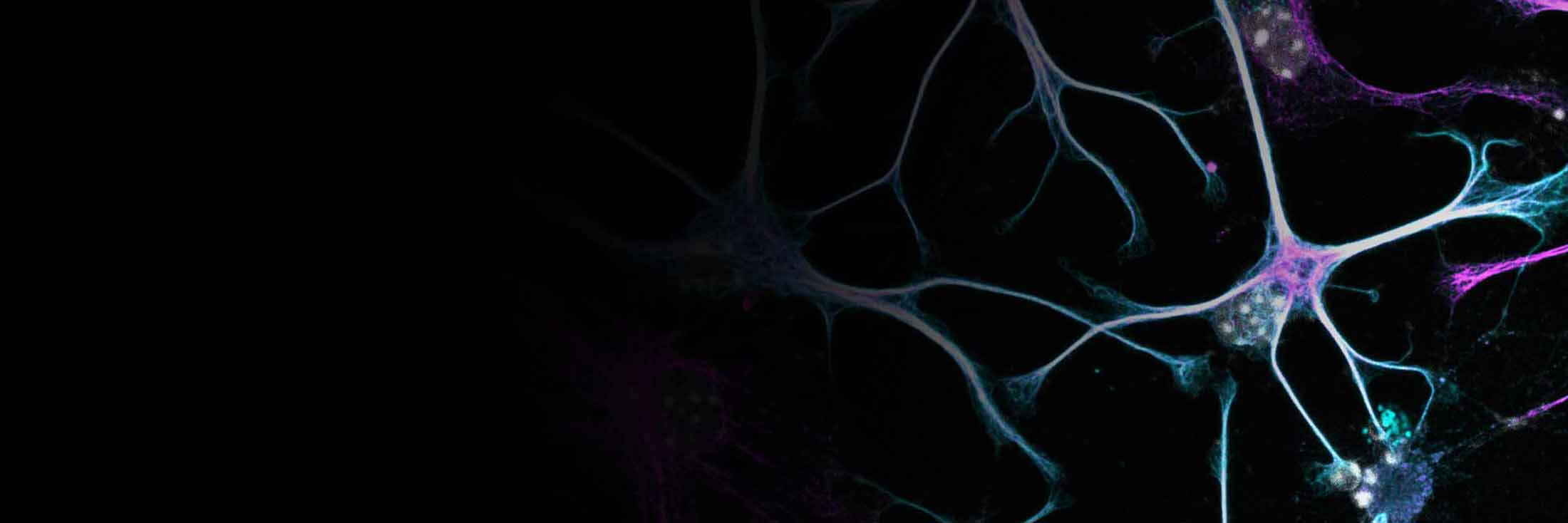
Using the developed techniques they have to date investigated everything from isolated cellular components and cell cultures to whole living tissues. An active research area of Kareem’s group is also working on better understanding the often quite unusual physical properties of biofluids, like blood and cerebrospinal fluid, and how these might be perturbed in pathologies. He and his team are particularly excited to look at less frequently studied biological and bio-relevant samples to see how the measured physical properties can give us a better understanding of function across spatial and temporal scales.
Brillouin Light Scattering Microscopy – a technique to look at mechanical properties
One technique at the heart of Kareem’s lab is Brillouin Light Scattering (BLS) Microscopy, an optical, non-destructive and label-free technique that allows for the characterization of physical properties and structure in biological samples via the speed and attenuation of hypersonic sound waves (so-called acoustic phonons) on the microscopic scale. “With BLS we can obtain insight into the mechanical properties at spatial and temporal scales that are inaccessible to other techniques, yet play a key underlying role for biological processes,” Kareem explains. “We are talking roughly about the characteristic time scales that water interacts with stuff (polymers, proteins,…), which can dictate phase and structural transitions. These can also affect macroscopic rheological/viscoelastic parameters that are essential for cell and tissue function which we can also measure in our labs.” As a condensed matter physicist by training, looking for and trying to make sense of exotic and dynamic material states that exist in biomatter and how these can make-or-break life, is evidently very exciting for him. He continues “When interpreted correctly, it can in some cases offer (part of) a bridge between molecular and collective (“mechanical”) properties, a connection between structural biology and cell biology if you will, that is tricky to both theoretically and experimentally get at, but essential for understanding life in all its complexity.”
Asked on the medical relevance he replies: “Inside a living cell things are continuously going through liquid-liquid and liquid-solid transitions, the latter not always being isotropic and responsible for the shapes of things. Insight into their dynamics, spatial correlations, symmetry properties, and so on, is important for understanding their origins and purposes, and ultimately being able to control them when they are not doing what they should be doing, as may be the case in certain diseases”. Such exotic structural/phase changes and their dynamics were recently shown by him and his team to impact cell wall growth in plants as well as DNA condensation and nuclear dynamics in mammalian cells (you can read more here). Kareem goes on “…this is of course all very fundamental and describes the ultimate goal, but in some cases the observation of significant correlations between a certain medical condition and the deviations of one measured parameter, can already motivate us to look into the translational potential–even if we are still figuring all the nitty-gritty details of everything that could be causing it”.
His excitement is palpable as he shows us the technologies in his lab. With their latest BLS spectrometers he and his team have studied a wide range of systems, spanning cell walls and cell nuclei, to muscle & nerve fibers, to teeth and fish guts.

A range of Brillouin Light Microscopy set-ups to support scientific exploration
Depending on the sample and questions being addressed the experimental setup will differ, and different BLS setups in the lab will be more or less suitable. For example, one setup that includes an imaging spectrometer, is best suited for studying live samples with a high spatial resolution. “This one is reasonably fast and not so invasive, so you could get a small image quite quickly to see how things spatially change over the course of say minutes or hours” explains Kareem. He mentions the setup is often used for various collaboration projects.
For higher temporal resolution and for processes that can be periodically stimulated, there is a setup with a special camera, to look at much faster “transient” changes in the mechanics that they are working on. Next, he shows us a setup that, while not so fast, has an extremely high spectral resolution and can be used to also measure the shear viscoelastic properties of non-transparent solid samples (relevant for certain processes). This is followed by a special detection configuration for measuring BLS at different scattering angles, which he mentions can also give insight into a material’s dispersion. “In the end it all depends on what you want to find out, and like everything, there is going to be some kind of compromise between spatial, temporal and spectral resolution, as well as the properties of the sample, that you will be forced to make” he mentions in passing.
Spotlight on clinical collaborations
For clinical collaborations, he also has a setup in the hospital, that makes it possible to measure samples immediately after being taken from patients. “In our collaboration with the hospital, we recently used both conventional shear rheology and BLS spectroscopy to study blood plasma’s viscoelastic and rheological properties in healthy samples compared to those of COVID-19 patients with different disease severities and outcomes” explains Kareem. The former technique here measures the shear properties on relatively speaking long time scales, while the latter measures the acoustic damping properties at picosecond-scale times which are acutely sensitive to the nature of intermolecular interactions and chemistry as well as being related to the microscopic viscosity. Taken together they paint a more complete picture of the physical-chemical properties of blood plasma. “That study in a way also demonstrated the potential medical usefulness of the BLS spectroscopy -measured parameters of blood plasma for rapid medical diagnostics, although there is still a bit of work to do before you’ll see it in your doctor’s office” he adds. (You can read more on this project here). At the hospital they are now looking at the effect of other pathologies on the BLS measured parameters of blood components that may have clinical translational potential, while in his labs at the Division of Anatomy they are also busy working on the ambitious task of assembling a “Brillouin Light Scattering Atlas” of the whole human body. The goal here is on the one hand to see in what other tissues it could have diagnostic or prognostic value, but it should also serve as a useful reference for other laboratories around the world employing the technique and groups wanting to use the measured values in biophysical modelling and simulations. Kareem explains this as “…it is kind of like a surveillance project, a first attempt at mapping what all is out there, that we and others can then use as a reference to build on. Creating some kind of map is usually a good starting point, regardless of where you want to eventually go.”
Potential for correlative workflow
BLS spectroscopy measurements can readily be correlated with other optical microscopy measurements (such as fluorescence and Raman confocal microscopy or even fluorescence correlation spectroscopy – something the lab has also been working on). It can also be correlated to other techniques probing mechanics on different scales, and the lab has some experience correlating BLS data with Atomic Force Microscopy (AFM), as demonstrated with e.g. a group in the Czech Republic for measuring the mechanical properties of cell walls (Link to the original work) and groups in Vienna studying collagen and muscle fibrils (Link to the original work).
Kareem warns however that “while it may seem natural to compare mechanical properties measured using BLS with those measured using other techniques, and from an experimental standpoint it might not be all too challenging to do, interpreting such data is everything but trivial given the different mechanics being measured. One really needs to think about this on a sample specific basis, as for example AFM and BLS can tell you two quite different things that may or may not be related”.
His lab is also developing and applying instruments to compress and stretch biological samples (including a Langmuir-Blodgett Trough microscopy setup for artificial lipid layers), which combined with the optical-probing techniques allow for the study of a materials’ non-linear microscopic viscoelastic properties in response to external stresses and finite strain.
While Kareem is open to all collaborations, the lab is currently particularly interested in collaborations that involve (1) incorporating measured parameters in multi-scale models/simulations to describe bio-relevant processes; (2) correlating results with those of techniques that can measure changes in chemistry or other physical properties on the sub-micron scale and on short time scales (sub-microsecond – millisecond scale); (3) biological systems where based on the physical-chemical changes there is a clear potential for medical translation; (4) applications that can give fundamental insight into the emergence of collective phenomena on sub-cellular scales.
So, how can you collaborate?
Interested researchers should reach out via the Austrian BioImaging or Euro-BioImaging portal. Together, you will discuss the project, decide on the goals, whether studies are feasible/make sense, and then what instruments would work best. Kareem explains “We usually perform experiments together with the researchers since instruments are not your simple plug-and-play type microscopes. Typically, data analysis is done in the lab, and data/results are uploaded to the cloud or shared via some other secured channel (depending on how big they are). We then discuss together what they can or cannot tell us”. Lastly, given that the labs are based at the Division of Anatomy and in close proximity to the hospital, the potential availability of human samples might be particularly interesting for some collaborators. “From a practical perspective we can, subject to all the necessary ethical approval, have access to diverse human tissue samples, but this of course needs to be discussed on a case-per-case basis and accordingly planned” remarks Kareem.

Magdalena Schindler
Published:
11 October 2024
Read the article as a PDF