An interview with Orestis G. Andriotis
A series of Euro-BioImaging Ambassador program – Introducing the Austrian BioImaging/CMI Node.
If you want to understand the physical properties of biological samples, Euro-BioImaging’s Austria BioImaging/CMI Node in Vienna might be a useful contact point for your research. Orestis G. Andriotis, a senior scientist in the group of Prof. Philipp Thurner at the TU Wien, has more than 10 years of experience in the Atomic Force Microscopy (AFM). As Ambassador of Euro-BioImaging, Magdalena Schindler recently got the chance to talk to him about his research motivations and his experience developing and combining different imaging modalities to probe the role of mechanical properties in biological contexts.
A multimodal imaging approach to study the architecture of life

Orestis (middle) is talking to Lena (left) and Baubak Bajoghli (right)
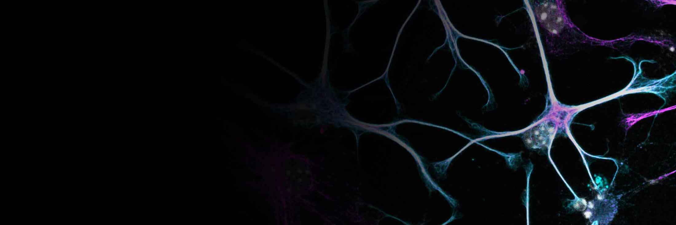
Collagen is one of the most abundant human proteins, giving structure to a multitude of tissues including ligaments, fascia and tendons. Each of those tissues needs different mechanical properties to fulfill their respective functions, but each is made at the ground substance level from the same component – the collagen fibril. Orestis G. Andriotis, a senior scientist in the group of Prof. Philipp Thurner at the TU Wien (formerly Vienna University of Technology), uses a range of multimodal imaging approaches to explore the versatility of collagen. As Ambassador of Euro-BioImaging, I recently got the chance to talk to him about his research motivations and his experience developing and combining different imaging modalities to probe the role of mechanical properties in biological contexts. Access to imaging techniques and his group’s expertise are available via Euro-BioImaging’s Austrian BioImaging/CMI Node.
Orestis is working at the Biomechanics and Mechanobiology Lab (B&M Lab) of TU Wien, where they work with different techniques such as atomic force microscopy (AFM), micro-computed tomography, micromechanical testing, and light microscopy. He has more than 10 years experience in the field, and still vividly remembers his initial drive for biomechanics. “In the beginning, it was the technicalities of it. When I was doing my PhD, I would need time to get trained for the instruments. Now that I have expertise in this, I know what goes wrong and what not. I can resolve it.” These days, his motivation has shifted to the biological questions. “The unknown, that’s what makes a project exciting for me now.”
Understanding the role of collagen in living systems with Atomic Force Microscopy
Although collagen is the primary structural component in all tissues responsible for mechanical function, talking to Orestis it becomes clear how unexplored the role of collagen fibrils is in many systems. “How do, for example, collagen fiber mechanics change in diseases and aging?” And it’s not only their amounts that matter to a healthy body, but also their molecular crosslinks and the feedback they give to the embedded cells. As a scaffold of the body, collagen plays a role in many diseases that are reflected in Orestis’ and his team colleagues’ projects, ranging from cardiomyopathy to pulmonary fibrosis and joint problems.
To answer his many questions, Orestis uses AFM, which employs a small cantilever to probe the deformability of a sample. This probing is done at high spatial resolution, resulting in a map of the mechanical properties of the sample. Orestis explains: “The atomic force microscopy is not destructive. So you push the sample only on the very surface.” This allows AFM to be used in series with other techniques, correlating different properties at the same spot in a sample. Orestis is collaborating, for example, with the group of Martina Marchetti-Deschmann, also a member of the Austrian BioImaging Node, to spatially map mechanical along with chemical properties acquired through matrix-assisted laser desorption/ionization (MALDI). This correlative multimodal imaging approach is not only applicable for fundamental research but has also proven useful in characterizing menisci from patients who underwent knee surgery.
Enriching the observations with other imaging approaches
Orestis further explains that AFM can even be combined with other measurements at the time of acquisition. “Together with Gerhard Schuetz (from TU Wien) we perform AFM with super-resolution fluorescence microscopy to look at the enzymatic digestion or the kinetics of the enzymatic digestion of these fibrils with time. We test spatially and temporarily how fibrils get digested at the nanoscale.” To further deepen our understanding of the physical properties of collagen fibrils, Orestis has explored, in collaboration with Gerhard Schuetz, a correlation with Stochastic Optical Reconstruction Microscopy (STORM). “With AFM, the problem is that you look at the surface. In other words, the AFM cannot image a part of the fibril that faces the sample holder. So with the STORM, what we could do is, we managed to image through the collagen-fibril and then we get a 3D cloud of the points that is now the structure of the fibril.”
A collaborative approach
How collagen fibrils change throughout our lifetime is a fundamental question that Orestis and the group of Philipp Thurner addressed in a collaborative project with Kareem Elsayad, a group leader at the Medical University of Vienna and a member of the Austrian BioImaging/CMI Node. In this project, they combined AFM and Brillouin light scattering microscopy to measure the effect of aging-characteristic modifications on collagen fibril hydration and their related mechanical properties. The outcome of this project was published in the Journal of Biomedical Optics Express in 2019.
The combination possibilities with AFM seem endless as Orestis guides me around the new lab space, which boasts plenty of room for new instruments and growth. He and Phillip Thurner are open for new collaborative projects. They have also good experience with sample transfer. For example, a group in Italy, flash freezes samples for Orestis to thaw in Vienna for AFM measurements.
Interested in collaborating?
If AFM could be useful for your research, many possibilities are available. Don’t hesitate to reach out to Austrian BioImaging/CMI or Euro-BioImaging for support.

Magdalena Schindler
Published:
11 October 2024
Read the article as PDF
