An interview with Gabriel Krens
A series of Euro-BioImaging Ambassador program – Introducing the Austrian BioImaging/CMI Node.
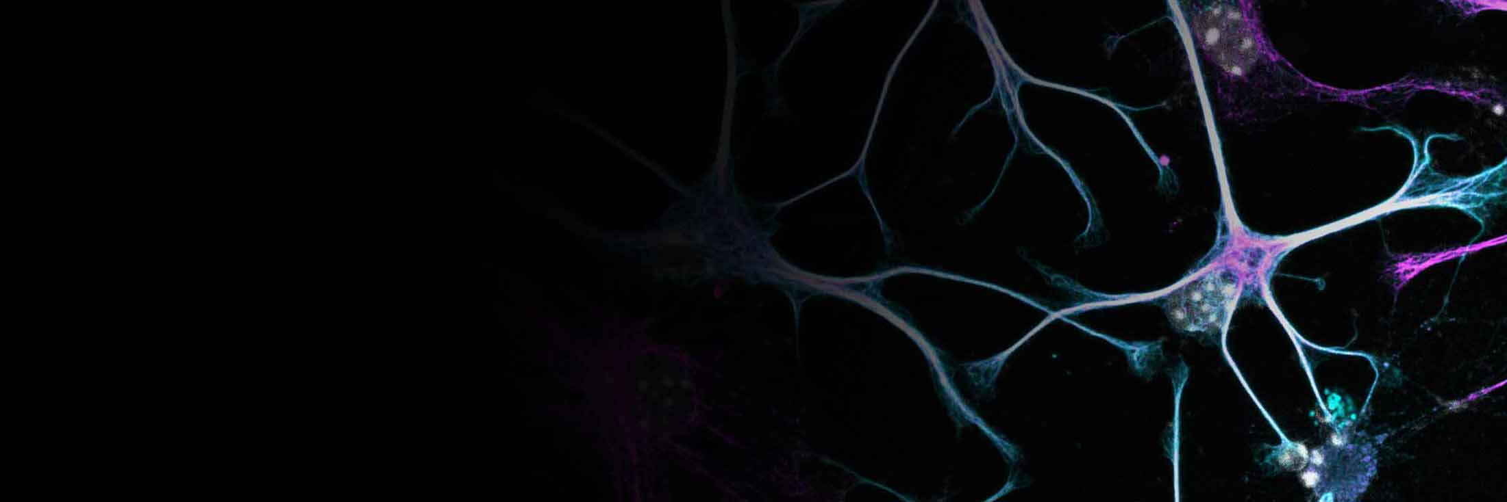
Seeing the European imaging infrastructure from a core facilities’ perspective
As a Euro-BioImaging Ambassador, I recently had the privilege of visiting the imaging & Optics Facility (IOF) at the Institute of Science and Technology Austria (ISTA). This cutting-edge core facility houses several advanced imaging technologies, including confocal, multiphoton, spinning disc, TIRF, light-sheet, and force-probing microscopy – as well as cell-cytometry and image analysis services . The facility is also a member of Austrian BioImaging/CMI, a Euro-BioImaging Node. During my visit, I had the pleasure of meeting the staff members, which introduced the services they provide. I also talked to Gabriel Krens, the manager of the core facility. Given my research focus on mechanobiology at EMBL, we talked about the core facility, the force-probing services available at ISTA and his expectations from the Austrian and Euro-BioImaging networks.
ISTA is a relatively new institute in Austria, now 15 years old. You joined the institute as a postdoc in 2010, one year after its opening. How was the imaging core facility at that time, and how is it now?
In the beginning, our imaging core facility had many confocal-based microscopes, but also advanced Multi-photon microscopes and an AFM, laser ablation system and “dual-pipette aspiration setup”. Now, we have integrated more spinning discs and other imaging technologies with higher resolution. We have further focussed on the further automation and integration of assays that include force-probing measurements, light modulation, and microfluidics based assays into imaging systems. These efforts aim to stimulate research projects and allow researchers to be more creative.
Could you please provide an overview of your core facility and explain services you offer, especially for force-probing biological samples.
Our core facility has three main service pillars: cell cytometry, imaging analysis, and microscopy. We divide the available technologies into different levels from 0 to 3 depending on how much effort is needed to learn them. The force-probing technologies are generally rather difficult to learn, in part because these are build atop of an imaging technology. In our core facility, we have the ability to probe from the cellular to the whole organismal level using different techniques, including atomic force microscopy (AFM) and dual pipette aspiration (DPA) assay. For force-probing, it is also possible to apply manipulations using UV- or IR- based laser ablation at the cellular and subcellular level, i.e., within the cell, it is possible to cut the microtubules. We also have ongoing pilots with magnetic- and optical tweezers.
How accessible are your force-probing services to external users?
Our approach is to integrate force-probing devices to commercial imaging setups as “add-ons”. Although our instruments at the ISTA campus are in high demand, we are always happy to help researchers who lack access to such instruments. I think this is everyone’s responsibility in science.
The assays are quite challenging to perform, and simply having access to the instrument is not sufficient. Apart from technical complexities, each project requires extensive customization and adaptation.
Yes, this is true. Researchers normally learn to operate a microscope within two days through training sessions and hands-on practice. However, force-probing is much more complex. It can take up to several weeks to master the preparation of samples and to perform measurements accurately.
Have you had external users utilize your force-probing services?
Yes, we have had several external users for the AFM, laser ablation setup, and dual pipette aspiration assay. However, most of these cases were part of collaboration with research groups at ISTA. As a core facility, we primarily offer support on the technological platform. Still, each project requires adaptation to the specific biological sample and solid understanding of biophysics to ensure users can properly analyze the image data they collected.
Do you support users in analyzing the image data?
We are putting more emphasis on image analysis services in general, as data is getting larger, and analysis is becoming more complex. For force-probing applications, we support image analysis and feature extraction, basically until the data can be plotted, but modeling and interpreting of force probing data in corresponding biophysical models lies with the user.
While force-probing can be challenging to support remotely, does your facility offer remote access for imaging technologies?
We have the philosophy that users should collect their own data to understand how it was generated, but also to get a feeling for the process that is studied. It is also very important for us to be able to provide direct feedback on the approaches taken. Therefore, we generally do not offer remote access for imaging technologies.
Now that external access to imaging core facilities works, how will the large quantity of generated image data be managed, and what are your expectations from Austrian BioImaging or other networks in this context?
We should not underestimate the challenges associated with the growing data size of advanced imaging applications and the infrastructure needed for data transfer. This is a bottleneck. My desire would be to find a solution at the national or even European scale to facilitate data exchange. If you want to perform correlated multimodal imaging, how can you transfer terabyte image data from between facilities and be able to work on that?
For example, transferring data from an imaging core facility to a lab server can be difficult ,especially when external hard drives / portable data-carriers are not permitted, and no internet access is available. So, we also need to think about connecting local infrastructure to overarching ‘data-exchange solutions’ for collaborative research projects. Additionally, /17I hope for improved data management systems, to better annotate and manage image data.
But is it not correct that your facility has implemented a cloud system to transfer up to 0.5 TB of data for external users?
That is true. However, I think we need to advance these local solutions into more centralized national or European repository systems, where researchers can rent space for their imaging projects. This will facilitate cross-node collaborations, particularly in image analysis, where some nodes have more expertise than others.
My wish is, therefore, to have an infrastructure that supports the storage of image data until the researchers publish their results. While we are very advanced in imaging technologies, we are somewhat behind in the efficient transfer of image data. Microscopy is just one of the image-producing instruments. We don’t need to reinvent the wheel, as in other fields, like astronomy, deal with even larger correlative datasets. Maybe collaborating with organizations like the European Space Agency (ESA) could offer valuable insights and solutions for addressing these problems.
Very interesting point. Thank you for taking the time to do this interview.

Magdalena Schindler
Published:
17 October 2024
Read the article as PDF
